Laser guided mole mapping
Mole mapping has long been an integral part of my clinical practice for monitoring patients at high risk for skin cancer, particularly melanoma. Combining clinical imaging with high resolution dermoscopy images allows a much more accurate monitoring of moles for signs of change.
Over the years I have detected many cases of early melanoma in this way, which makes it an essential part of my clinical practice. With technology improving it is always exciting integrating the latest technology into my mole mapping practice.
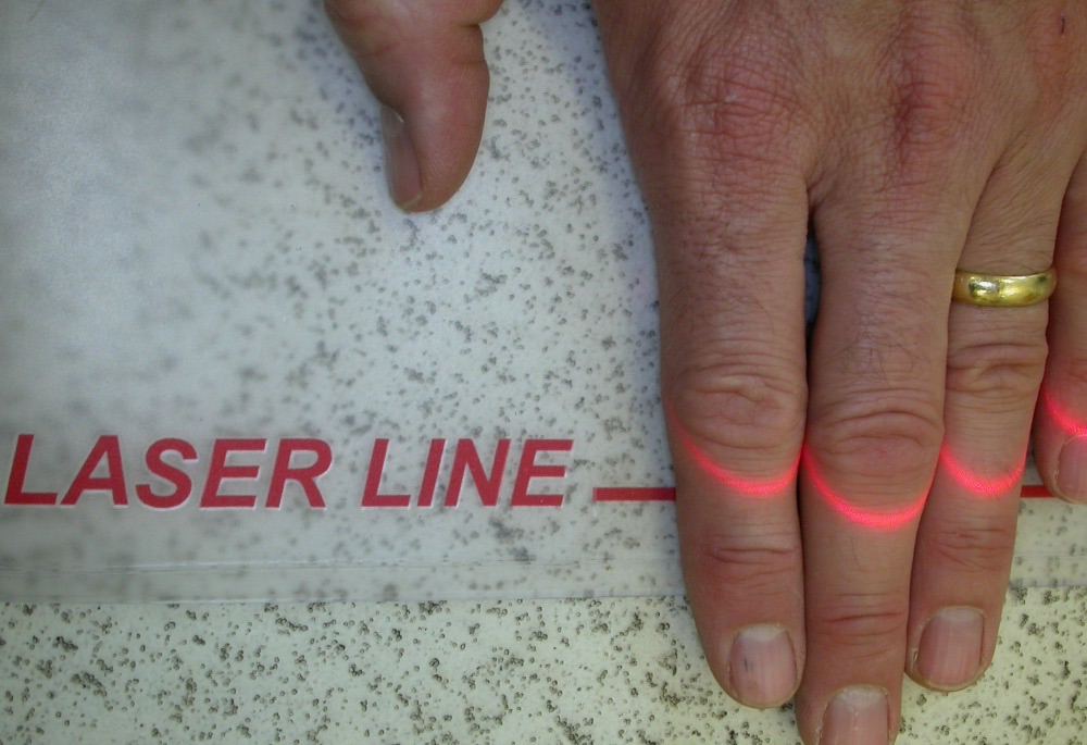
The latest addition to my skin cancer screening service includes a novel method of improving reproducibility of clinical imaging – laser guided mole mapping. Standardisation of placement of camera to patient is essential in promoting reproducible imaging. This system uses a fixed laser, mounted to the imaging system (FotoFinder) to help standardise foot placement during the mole mapping process. The result is a greater confidence in the quality of mole mapping, and thus better results.
What to expect at a mole mapping appointment
Dr Bowling’s mole mapping service comprises of three components.
Step 1. Dermoscopy assessment
Firstly, every patient gets a full dermoscopy examination of every mole, from the soles of their feet to the top of their scalp. This, dermoscopic mole screening, is essential in identifying the typical mole pattern for any individual. Once the typical mole pattern or mole patterns are known, a greater confidence and ability in finding the abnormal mole follows. Importantly, all forms of skin cancer are looked for in this method, such as basal cell carcinoma (BCC) and squamous cell carcinoma (SCC), and not just melanoma.
Step 2. Mole mapping
Secondly, if, following the dermoscopy assessment, the patterns of moles are more unpredictable or if you are at high risk for changes in moles, then mole mapping with a computerised imaging system may be considered. These clinical images provide a background template of your skin which is an excellent reference point for future surveillance.
Step 3. Digital dermoscopy
Finally, if there are a number of suspicious moles, detailed dermoscopic images can be taken. This is really helpful for any moles that have unpredictable dermoscopic features or a multicomponent pattern on dermoscopy. Images can be retaken in the future looking for any dermoscopic change, a feature often seen in early melanoma.
Take home message
For any mole mapping or mole screening service ensure that step 1 is performed by an expert, as it is typically at this stage that the most skin cancers are detected. Step 2 and step 3 will depend upon your risk factors for skin cancer.
Laser guided mole mapping is available in my Oxford clinic.
To book an appointment with Dr Bowling please call 01865 861577.

