What is dermoscopy?
Dermoscopy is a technique for examining the appearance of the skin – ordinary skin as well as moles – to diagnose skin problems. It consists of using a handheld device, dermatoscopes, which combines strong magnification with good lighting to +/- a polarising filter to enable your dermatologist to get the best view possible of your skin problem.
Although dermoscopy can be helpful in all forms of skin diagnosis, it is particularly helpful for the diagnosis of skin cancer, particularly melanoma.
Dermoscopy and moles
Dermoscopy can be used to zoom in on a mole and to accurately see diagnostic details under the surface which are otherwise invisible to the naked eye. Moles recorded by dermoscopy can be compared over time so that even the slightest change that might indicate a pre-cancerous or cancerous change can be detected.
Dermoscopy combined with digital photography produces accurate mole mapping and suspect moles are then examined by Dr Bowling again using a dermatoscope. If worrying signs are confirmed, the next step is to have a biopsy to rule out or confirm cancer.
Dermoscopy in action
Dermatoscopes are constantly evolving though tend to share the common features of bright LED illumination, x 10 magnification and polarising filters. Newer devices have UV lights which can provide further information for assessment:
Dr Bowling has many dermatoscopes available and uses them in addition to computerized digital mapping of moles, when necessary.
He is a recognised World expert in this technique.
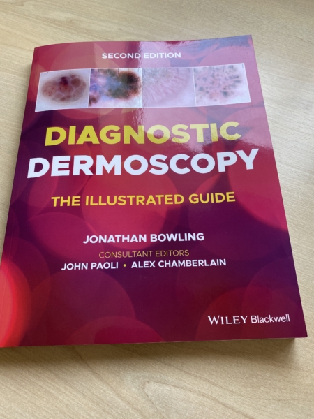
2nd Edition of textbook
_____________________________________________
“For more information on dermoscopy take a look at my textbook on dermoscopy, which is available in 8 languages and used Worldwide.”
____________________________